Let VAHEAT do the rest.
 Extended temperature range up to 200 °C
 Temperature stability of 0.01 °C (rms)
 Superb image quality
Key Features
VAHEAT is a precise temperature control unit for optical microscopes. It combines local heating with direct temperature sensing in the sample volume. This allows for fast and precise temperature adjustment with heating rates up to 100°C/s while maintaining highest temperature precision. Made for investigations of temperature-sensitive processes in life sciences and material research.

FAST &
RELIABLE
FAST &RELIABLE
VAHEAT lets you control the temperature inside the field of view independently from the type of microscope objective or the objective’s temperature. The system is designed as standalone unit without the need for any additional modifications to the optical setup (e.g objective heater) in order to avoid a temperature sink in your field of view. Additionally, the specific design of our smart substrates ensures that the objective’s performance is not altered even at higher temperatures.

EXTENDED TEMPERATURE
RANGE UP TO 200 °C
EXTENDED TEMPERATURE RANGE UP TO 200 °C
Extend your experimental temperature range to 100°C (standard range) or even up to 200°C (extended range) depending on your experimental needs. The standard range version is compatible with oil-immersion systems while the extended range version can be operated with air objectives.
HEATING
MODES
HEATING MODES
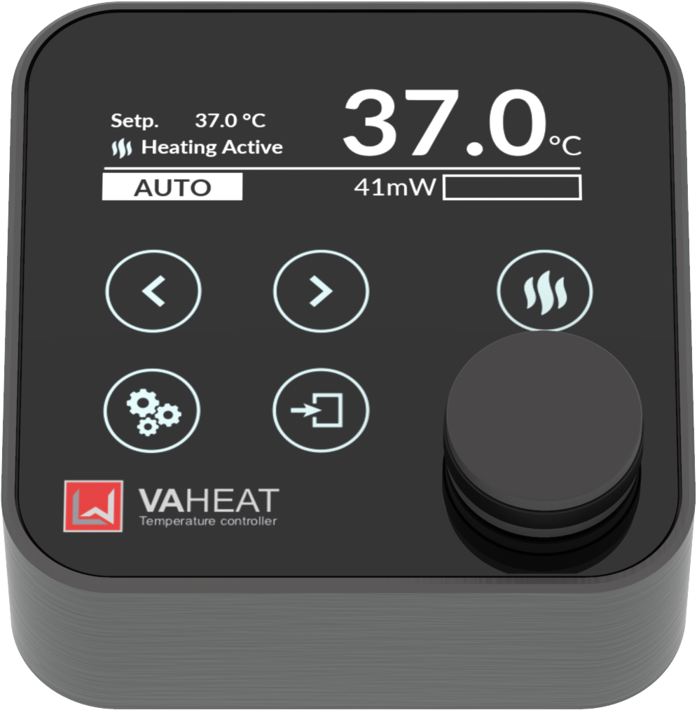
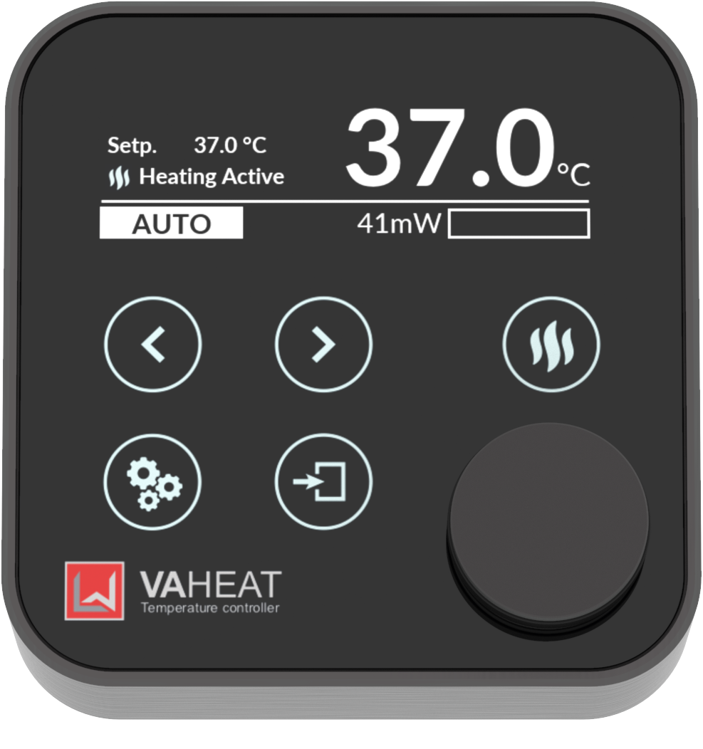
VAHEAT is equipped with four heating modes for different experiments depending on your needs. Modes for fast heating, auto-compensated heating, or well-defined temperature profiles are available.

SUPERB IMAGING
QUALITY
SUPERB IMAGING QUALITY
No optical aberration up to 80 °C with the highest numerical aperture objectives on the market. Perfectly suited for single molecule and super resolution studies using state-of- the-art methods (STORM, STED, TIRF, etc.). For more see Technical Performance.
MECHANICAL STABILITY &
DEVICE COMPATIBILITY
MECHANICAL STABILITY & DEVICE COMPATIBILITY
No thermal drifts or vibrations even at elevated temperature allow precise single molecule localization. We designed VAHEAT to be compatible with all commercial microscopes. No further modifications of your setup are needed. Its fast thermal response allows for nearly instantaneous thermalization tremendously reducing the waiting times as usual with conventional heating systems.

TEMPERATURE STABILITY
OF 0.01 °C (rms)
TEMPERATURE STABILITY OF 0.01 °C (rms)
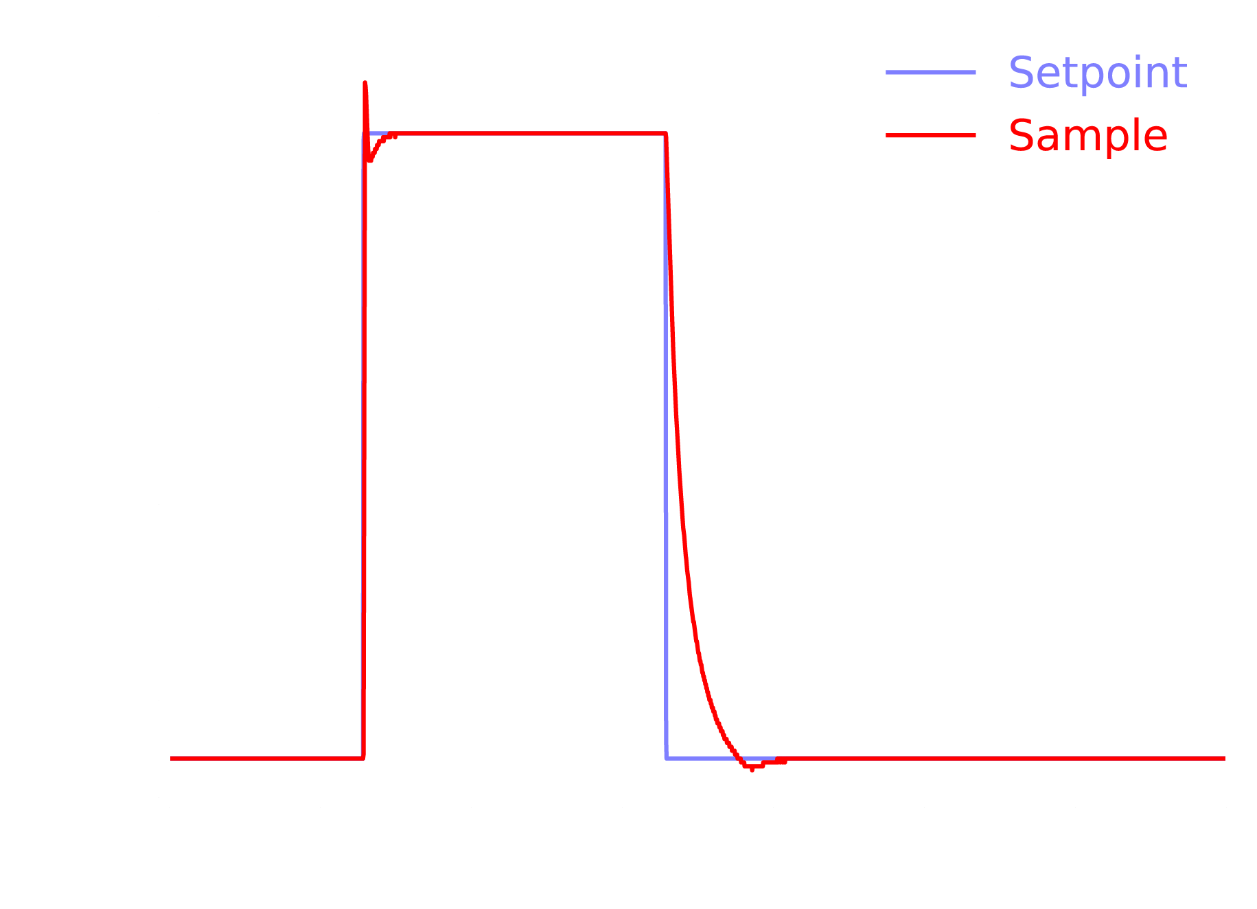
Extreme temperature stability on long (hours to days) and short (seconds to minutes) time scales down to 0.01°C (rms). External temperature variations due to air flow, fluid exchange etc. will be detected and compensated via direct temperature feedback inside the sample volume.
System Components
Smart Substrates
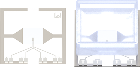
SMS-P
Dimension: 18x18x0.17 (± 0.05) mm³
Temp. range: RT - 105°C
The standard range smart substrates are optimized for high-resolution studies similar to #1.5 coverslips. They have a size of 18 x 18 mm² and a thickness of 170 µm. We specify their full functionality up to 100 °C with heating powers up to 2.5 W. Your sample can be mounted using reservoirs, flow chambers or even your microfluidic device.
SMS-E
Dimension: 18x18x0.5 mm³
Temp. range: RT - 200°C
The extended range smart substrates are made for temperatures up to 200 °C. They are 500 µm thick, similar to #5 coverslips and are compatible with the extended range control unit. Also here multiple configurations for mounting your sample are available.
We offer a variety of different substrates and options to mount your sample depending on your application. This includes larger heated areas, dual chamber reservoirs, flow chambers, and many more.
By answering application-specific questions we can point you towards the right smart substrate configuration! Just click here.
Microscope Adapter
The microscope adapter fits into most common microscope stages due to its size of 75 mm x 25 mm (3” x 1”). Its slim design with a maximal height of 11 mm allows unrestricted access from above. The microscope adapter is thermally insulated from the heated area and stays at room temperature even for sample temperatures of 200 °C.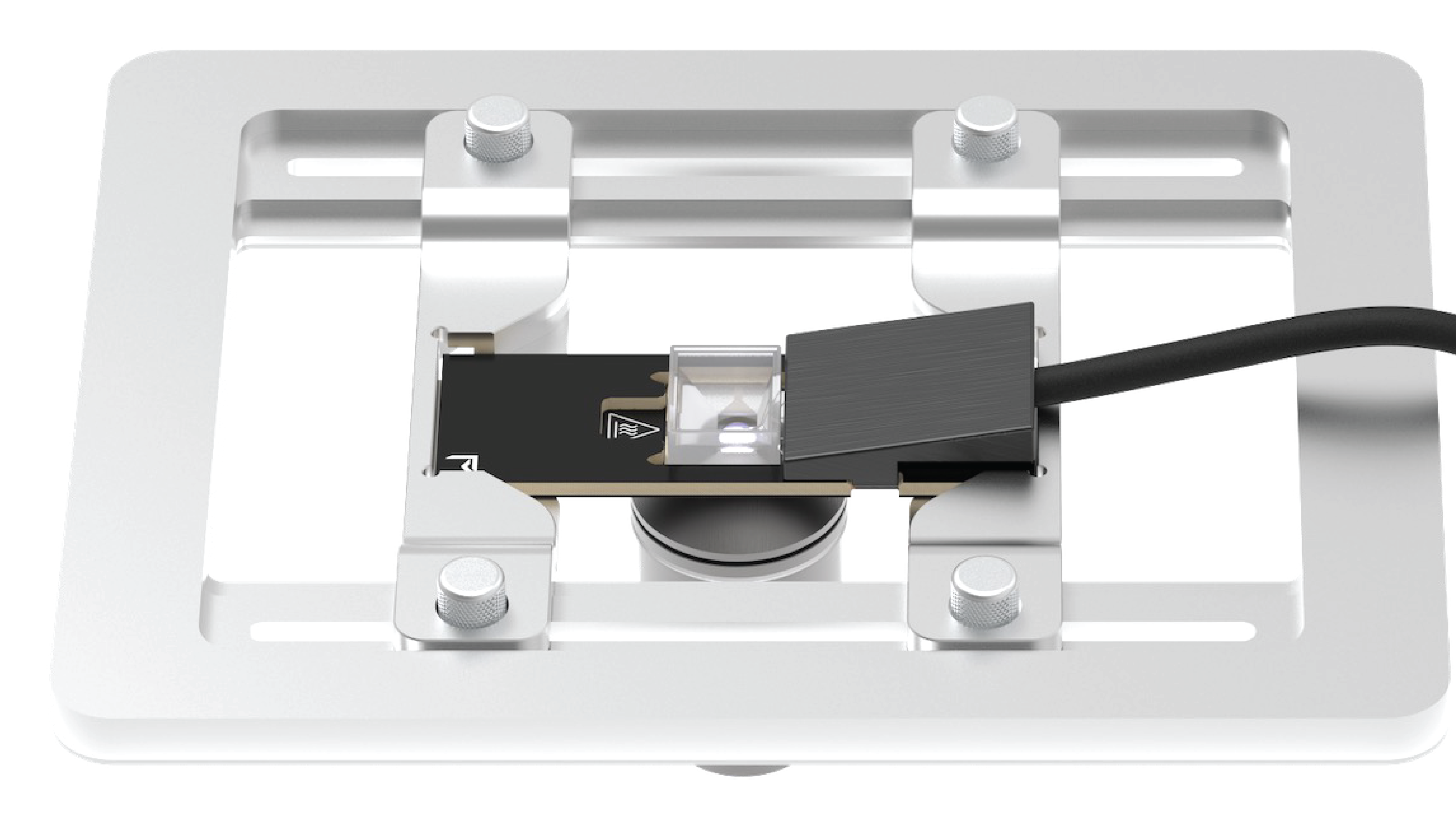
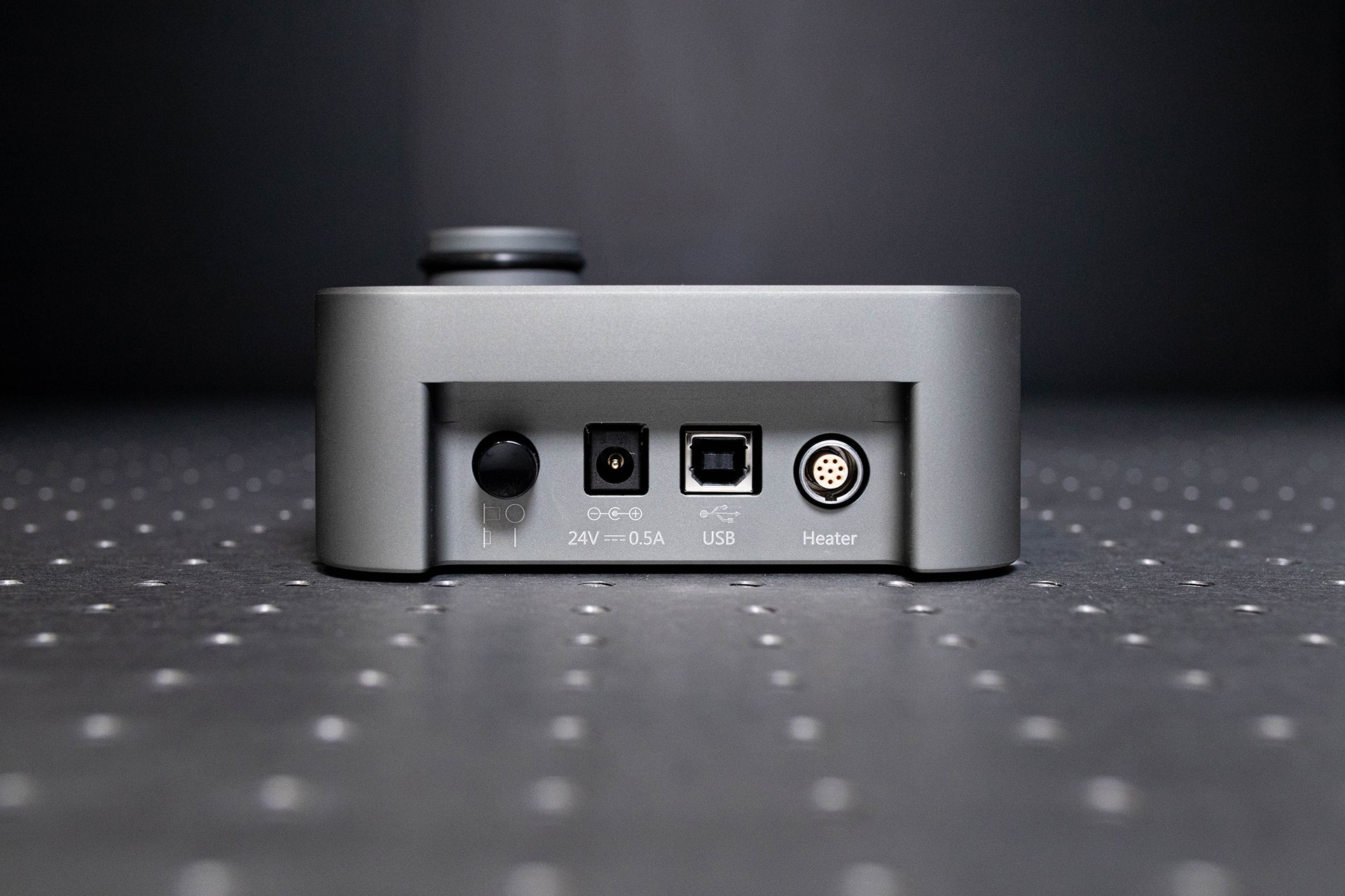
Control Unit
Extended Range
- <5000 mW
Heating Power
- 200°C
Max. Temperature
 SmS, SmS-R & SmS-E
Smart Substrates
- High Resolution Studies
Applications
Standard Range
- <2500 mW
Heating Power
 105°C
105°C
Max. Temperature
 SmS & SmS-R
Smart Substrates
 High and Super Resolution Studies
Applications
This is what our Customers have to say

Dr. Connor Bischak
University of Utah

Prof. Hendrik Dietz
TU Munich

Dr. Kerstin Göpfrich
MPI for Medical Research, Heidelberg

Dr. Alexandre Bisson
Brandeis University

Dr. Senthil Arumugam
EMBL Australia/Monash University

Prof. Laura Kaufman
Columbia University, New York City
Dr. Liz Birchall
University of Nottingham

Dr. Marleen van Wolferen
University of Freiburg

Serena Teora
Radboud University

Dr. Josef Gotzmann
Head of BioOptics Facility, Max Perutz labs

Tobias Martens
Cell Imaging Core at KU Leuven

Dr. Bas van Ravensteijn
Eindhoven University of Technology (Currently: Utrecht University)

Dr. Tugce Oz Yoldas
MPI of Biochemistry, Martinsried

Dr. Linda Rubio
LSU Health, Shreveport
Technical Performance
100x, NA=1.46, oil immersion objective
40x, NA=0.4, air objective
The imaging quality at elevated temperature up to 80°C is not altered using VAHEAT even when working with the highest numerical aperture objectives commercially available. To prove this, we experimentally determined the three-dimensional point spread function for various sample temperatures. The x-y resolution only increases slightly at temperature above 80°C due to increased particle diffusion. An elongation of the focus in z-direction arises with increasing temperature due to the temperature dependence of the immersion oil. This effect strongly depends on the material properties of the immersion medium and can be compensated by considering an effective change of optical path length. For air-spaced objectives the imaging quality is not affected.
Compatible Imaging Techniques
 Total internal reflection microscopy (TIRM)
- Atomic force microscopy (AFM)
- Confocal microscopy
- Super resolution methods (SIM, STORM, PALM, PAINT, STED)
- Interferometric scattering microscopy (iSCAT)
- Total internal reflection microscopy (TIRM)
Application Examples
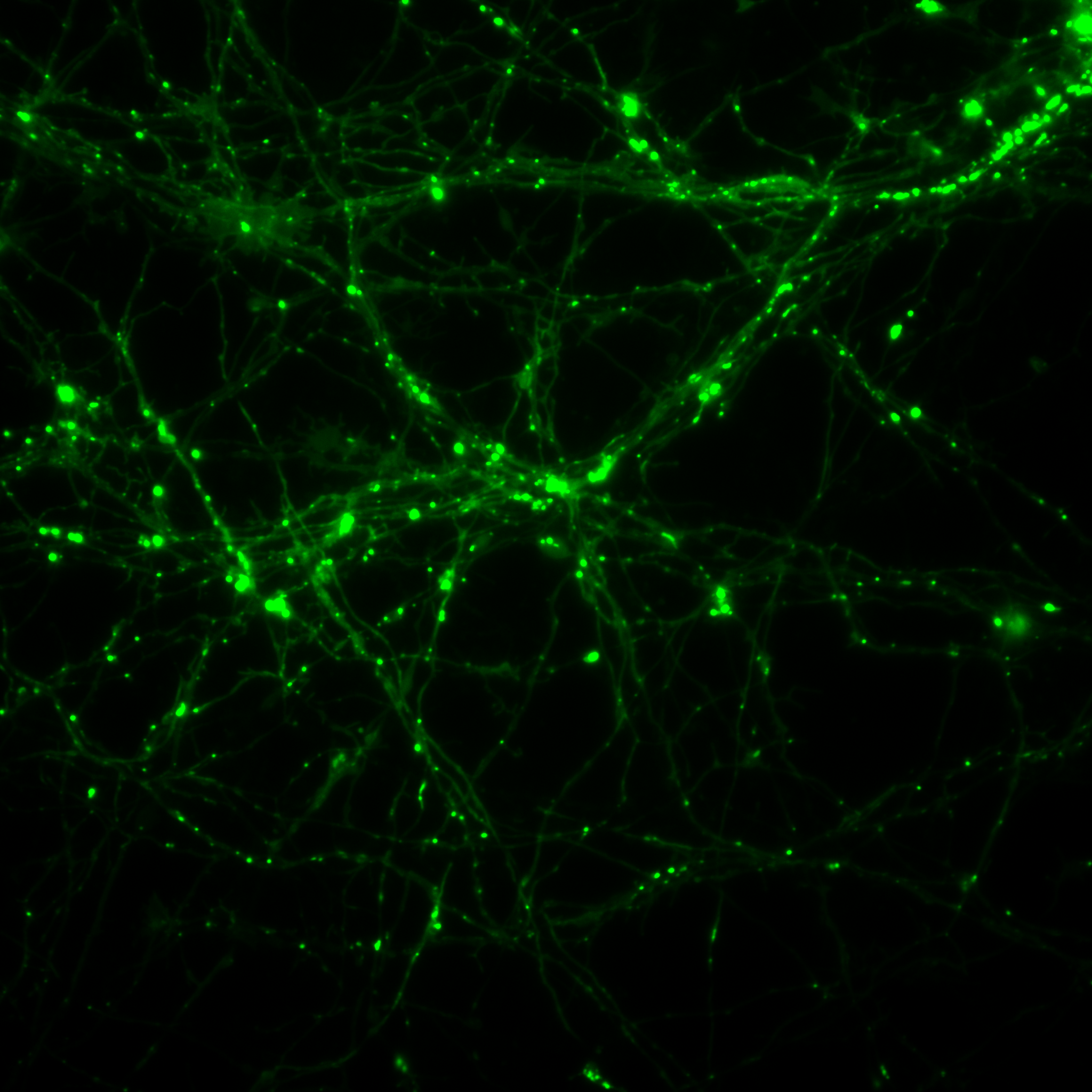
Living organisms are highly sensitive to changes in their environmental conditions, especially to temperature. VAHEAT ensures reliable and precise temperature control while transfer and during imaging. Investigating temperature sensitive processes of cellular behavior, such as Ca2+ activity in multicellular tumor spheroids or thermal stimulation of neurons has never been this easy.
Exploit VAHEAT ‘s capabilities as micro stage top incubator.
CATEGORIES:Cell Biology | Neuroscience | Medicine | Stage Top Incubator